News

暗场成像+SAXS:纳米材料微观探测新路径

By 2025-11-12 No Comments

融合成像与散射技术,追溯暗场信号的起源

X射线暗场(DF)成像可提供样品内纳米级和微米级结构异质性的存在及分布信息。这些异质性包括纳米孔隙、微裂纹或界面等,在传统成像中难以分辨。与依赖吸收衬度且受探测器分辨率限制的传统X射线成像不同,暗场成像能呈现小于像素尺寸的结构。其原理是通过探测X射线散射的局部变化:当X射线与未分辨结构相互作用时,会发生小角度偏转。这些散射事件的强度和空间分布可被绘制成图,形成暗场图像。

获取X射线暗场(DF)图像的实验方法与装置配置有多种。这些技术最初在同步辐射装置中开发,核心是利用结构化掩模或光栅等光学元件,在样品前端对X射线光束进行调制。首先仅用调制光束记录参考图像,随后将样品置于光路中拍摄第二幅图像。与参考图案相比,样品会导致调制信号出现局部模糊或可见度下降。通过数值量化这种模糊效应,即可重建暗场图像(见图1)。在实验室装置中,这一原理可通过模基调制成像(MoBI)方法 [1] 实现,该方法也是Xeuss Pro系统暗场相衬成像(DF-PCI)功能的核心基础。

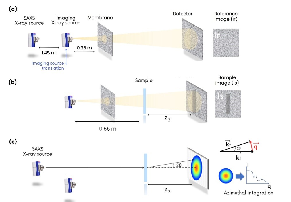
图1. Xeuss Pro系统上的暗场成像-SAXS联用装置示意图。该装置通过一片特殊膜层调制X射线光束,从而生成相位衬度(a,b)与暗场衬度图像;同时,SAXS测量(c)则记录来自同一样品区域的散射强度I(q)。这种独特的配置,首次实现了空间分辨的暗场图像与SAXS散射曲线的直接关联。

Correlating X-ray Dark-Field Imaging with SAXS Measurements to Probe Nanostructured Materials

To download the application note, please fill in this form. You will receive a link by email immediatly.

My main areas of interest*
Nanoparticles and colloids
Biostructural research
Polymer research
Food science
Cosmetics and consumer care
Renewable energy and storage
Inorganic materials
Semiconductors
Pharmaceuticals
Other
I would like to discuss about the SAXS technique or SAXS instruments*
Yes
No
I have a SAXS instrument purchase project*
Yes
No
Privacy*
By clicking this button you agree that Xenocs collects and uses your personal data to send you information relevant to your areas of interest. For more information, please visit our Privacy policy page https://www.xenocs.com/privacy-policy/

目前学界普遍认同,暗场信号由亚分辨率结构引发的X射线散射产生。但这些散射事件的具体本质,以及信号的角度灵敏度,仍是当前的研究热点。最简单的模型将暗场信号的起源仅归因于样品内部的多次折射现象 [2, 3]。与之相反,更复杂的模型则尝试将暗场测量事件与小角X射线散射强度进行直接关联 [4, 5]

在本应用手册中,你将了解Xeuss Pro如何将暗场成像的空间定位能力,与SAXS的统计解析能力合二为一,为纳米尺度结构研究提供互补性见解。它的独特之处在于:您可以在同一位置、无需移动样品的情况下,同步获得散射强度与空间分布信息,实现两种数据的完美关联。

本研究运用联用技术,成功揭示了暗场信号的物理起源:究竟是哪些散射事件和角度范围,主导了最终的图像衬度。通过对比相同材料的暗场图像与SAXS曲线,我们证实多次折射与小角散射共同塑造了暗场衬度,并且,其信号灵敏度主要覆盖在低q值区(USAXS)。

本研究结果源自更系统性的研究,完整方法论与分析详见文献[6]。

Author: C. Magnin
Measurements and Data Analysis: C. Magnin
Scientific reviewer: A. Tutueanu.
[1] C. Magnin, L. Quenot, D. M. Cenda, B. Lantz, B. Faure, and E. Brun. On the optimisation of the geometric pattern for structured illumination based X-ray phase contrast and dark field imaging: A simulation study and its experimental validation. (2024). DOI: 10.48550/arXiv.2504.10665
[2]R. von Nardroff. Refraction of X-Rays by Small Particles. Physical Review 28, 240 (1926). DOI: 10.1103/PhysRev.28.240
[3] Y. Y. How and K. S. Morgan. Quantifying the X-ray Dark-Field Signal in Single-Grid Imaging. Optics Express 30(7), 10899–10918 (2022). DOI: 10.1364/OE.451834
[4] M. Strobl. General Solution for Quantitative Dark-Field Contrast Imaging with Grating Interferometers. Scientific Reports 4, 7243 (2014). DOI: 10.1038/srep07243
[5] P. Modregger, M. Kagias, S. C. Irvine, R. Brönnimann, K. Jefimovs, M. Endrizzi, and A. Olivo. Interpretation and Utility of the Moments of Small-Angle X-ray Scattering Distributions. Physical Review Letters 118, 265501 (2017). DOI: 10.1103/PhysRevLett.118.265501
[6] C. Magnin, M. Fernández Martínez, D. M. Cenda, B. Lantz, S. Barton, B. Faure and E. Brun, How X-ray dark-field imaging relates to small-angle X-ray scattering measurements, Applied Crystallography, 58(4), (2025). DOI: 10.1107/S1600576725004017